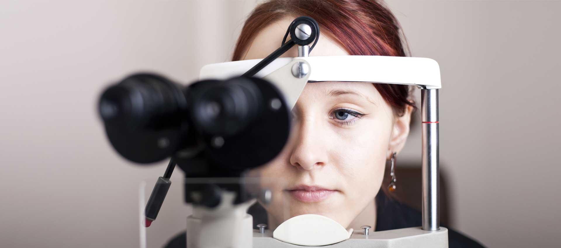

Diabetic Retinopathy detection requires capturing a photograph of the retina using specialized equipment such as a slit-lamp and fundus camera. The image is then examined by an ophthalmologist, optometrist or a trained professional to detect abnormalities such as microaneurysms, exudates, hemorrhages, macular edema, etc. to determine if DR is present and its severity and stage of progression. In general DR can be classified as mild, moderate or vision-threatening, which includes severe non-proliferative DR, proliferative DR (PDR) and diabetic macular edema (DME). Accurate diagnosis of DR from fundus camera images and grading its severity requires professional expertise and training.
In recent years, many AI systems using deep learning have been very successful in image recognition and classification tasks. For example, in the Imagenet challenge, requiring identification of objects in a 1000 categories, the best models achieve a classification error rate of less than 5%. – exceeding the best human accuracy levels

Many of these models, have now been adapted successfully for use in a variety of medical image diagnosis tasks such as melanoma, breast, lung cancer detection and diabetic retinopathy.
In particular, a team at Google published results in 2016 of a study for detecting DR working with doctors in India and the US. The results show that their AI model’s performance for DR detection and grading its severity was on-par with that of ophthalmologists. Their model had a combined accuracy score of 0.95, which was slightly better than the median of the 8 ophthalmologists consulted (measured at 0.91).